 By
Supporters of two rival theories of consciousness agreed in advance how an experiment could prove them wrong, had theory-neutral teams run it on 256 people, and found that neither theory's central prediction held in full. The result appeared in Nature on 30 April 2025 with both camps among the authors.
A group called the Cogitate Consortium recorded brain activity in laboratories in the United States, Britain, the Netherlands and China while volunteers looked at pictures of faces, objects, letters and made-up symbols. More than 40 researchers signed the paper, including Christof Koch and Giulio Tononi, who back integrated information theory, and Stanislas Dehaene, a leading proponent of global neuronal workspace theory.
Michael Pitts, a professor of psychology at Reed College, co-designed the experiments and has co-led Cogitate since 2019. A co-senior author of the 2025 Cogitate paper in Nature, he said in an email: "Overall, I was surprised to see how many predictions made by both theories turned out to be supported by the data."
The two theories disagree about where in the brain a conscious experience arises. Integrated information theory, known as IIT, "suggests that consciousness emerges when information inside a system (like the brain) is highly connected and unified", Koch, a meritorious investigator at the Allen Institute in Seattle, told GeekWire. Its backers place that unified network in a "hot zone" at the back of the brain, where the temporal, parietal and occipital lobes meet.
Global neuronal workspace theory, or GNWT, holds that a piece of information becomes conscious when it is broadcast across the brain, and that the prefrontal cortex, the region behind the forehead, plays the central role in that broadcasting. The theory predicts "ignition", a brief, sharp burst of prefrontal activity about a third to half a second after a stimulus appears, and a second burst when it disappears.
The agreement struck in advance, known as preregistration, meant the two sides wrote down, before any data were collected, what would be measured, what would count as a pass or a fail for each theory and how the results would be read. The paper says the proponents and the consortium "developed and preregistered the experimental design, divergent predictions, expected outcomes and interpretation thereof". Teams with no stake in either theory then gathered and analysed the data.
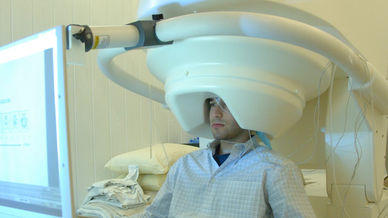
The consortium used three ways of watching the brain. Functional magnetic resonance imaging, or fMRI, tracks blood flow across the whole brain but updates only every second or two. Magnetoencephalography, or MEG, picks up the faint magnetic fields produced by electrical activity in neurons and records timing to the millisecond.
Intracranial recording uses electrodes placed inside the brain, here in 34 patients with drug-resistant epilepsy who already had electrodes implanted for clinical reasons, of whom 32 provided data after two were excluded for incomplete recordings. Of the 256 volunteers, 120 were scanned with fMRI and 102 with MEG.
Three predictions were tested. The first concerned decoding, which means training a computer program to work out from the brain signal alone what the person was looking at. IIT predicted decoding would work best from the back of the brain and that adding prefrontal signals would not improve it. GNWT predicted the prefrontal cortex would hold that information too.
Both were partly right. Whether the picture showed a face or an object could be decoded from the back of the brain and from the prefrontal cortex, in every recording method. The orientation of a face, whether it looked left, right or straight ahead, could be read from the back but not from the front in the intracranial and fMRI data, and only weakly in MEG.
"Here the evidence is decidedly in favor of the posterior cortex," Koch told GeekWire. "Either information pertaining to the conscious experience couldn't be found in the front, or it was far weaker than in the back."
Pitts, who uses the shorthand PFC for the prefrontal cortex, drew a more mixed lesson from the same test. Volunteers were told in each part of the experiment which images mattered for the task, he said, so "it was interesting to find evidence for decoding of some aspects of stimuli in PFC, even when they were irrelevant to the task, as predicted by GNWT".
He added: "It was also interesting to test the prediction made by IIT that adding PFC areas to posterior areas should not increase the accuracy of decoding (beyond posterior areas alone). This was indeed what the data ended up showing, although as we said from the outset, this result does not necessarily challenge GNWT."
The second prediction concerned time. Images stayed on screen for half a second, one second or one and a half seconds. IIT said activity at the back should last as long as the image did. GNWT said the prefrontal cortex should ignite at the start and again at the end, with a quiet gap between.
In the intracranial recordings, 25 electrodes at the back of the brain tracked how long the image was shown, mostly in early visual areas and the fusiform gyrus, a region involved in recognising faces. In the prefrontal cortex, 123 electrodes responded when an image appeared, 99 to any image and 24 to particular categories. None of the 655 prefrontal electrodes showed the burst at the start and again at the end that GNWT predicted, even though end-of-image responses were found in visual areas to the same images.
The third prediction concerned how regions talk to each other. IIT predicted sustained, high-frequency synchronisation within the back of the brain, linking early visual cortex to areas that recognise faces and objects. GNWT predicted brief long-range synchronisation between those recognition areas and the prefrontal cortex.
The preregistered measure supported neither theory. An exploratory follow-up found high-frequency synchronisation, in what is called the gamma band, between the prefrontal cortex and face- and object-selective areas inside the time window GNWT predicted, and the paper's summary reports "content-specific synchronization between frontal and early visual areas". The sustained link within the back of the brain that IIT required did not appear.
"The challenge is to come up with ways to explain the null result in the third prediction," Koch told GeekWire. "We're working on that."
The paper's own summary is that the results "align with some predictions of IIT and GNWT, while substantially challenging key tenets of both theories". It adds that the challenges extend to other theories that share some of the same predictions, including higher-order theories that place conscious content in the prefrontal cortex and recurrent processing theories that place it at the back.
The project began at a workshop at the Allen Institute in 2018 and was supported by the Templeton World Charity Foundation. A progress report in June 2023, delivered at the Association for the Scientific Study of Consciousness meeting in New York, found no clear winner.
That result cost Koch a bet he had made with the philosopher David Chalmers in a bar in Bremen in 1998, when he wagered that a specific brain signature of consciousness would be found within 25 years. He paid with a case of Portuguese wine, The Debrief reported.
Three months later, 124 scholars posted an open letter on the preprint server PsyArXiv arguing that IIT should be labelled pseudoscience. Nature reported at the time that the letter "has caused an uproar in the consciousness-research community". The Cogitate preprint was circulating as the argument ran, and the IIT side still signed the finished paper, including the section recording the predictions their theory failed.
Koch told GeekWire the stakes are practical as well as academic. He cited a study reporting that around a quarter of unresponsive patients are conscious but "unable to signal this at the bedside".
The authors list their own limits. Only 12 intracranial electrodes were located in the earliest visual areas, compared with 472 in the prefrontal cortex, in the patients used for the exploratory connectivity analysis, which weakens the test of IIT's connectivity claim. The 655 prefrontal electrodes mentioned earlier come from a different analysis, the search for start-and-end bursts across 31 patients. The authors also note that they cannot fully rule out task effects on category, that the study does not capture the full richness of experience, and that it has no recordings from single neurons.
Pitts said follow-up experiments on the failed predictions are under way. "I am most excited to see that subsequent experiments are already being conducted to follow-up on some of the failed predictions, such as the apparent lack of an offset signal in PFC."
Sources
- Cogitate Consortium, Adversarial testing of global neuronal workspace and integrated information theories of consciousness, Nature, 30 April 2025
- GeekWire, Study turns up new clues, new questions in the search for the roots of consciousness, 30 April 2025
- Nature news, Consciousness theory slammed as 'pseudoscience', 20 September 2023
- The Debrief, 25-Year Academic Wager Settled, 27 June 2023
- Michael Pitts, email to Justin Brown, 14 October 2026