 By
Heartbeats are followed by a measurable cortical response, recorded under the name heartbeat evoked potential, and the reference list of the study below includes work on it going back to the early 1990s.
In 17 volunteers, the size of that trace in the window just before a faint striped pattern appeared on a screen predicted whether the volunteer reported seeing the pattern at all.
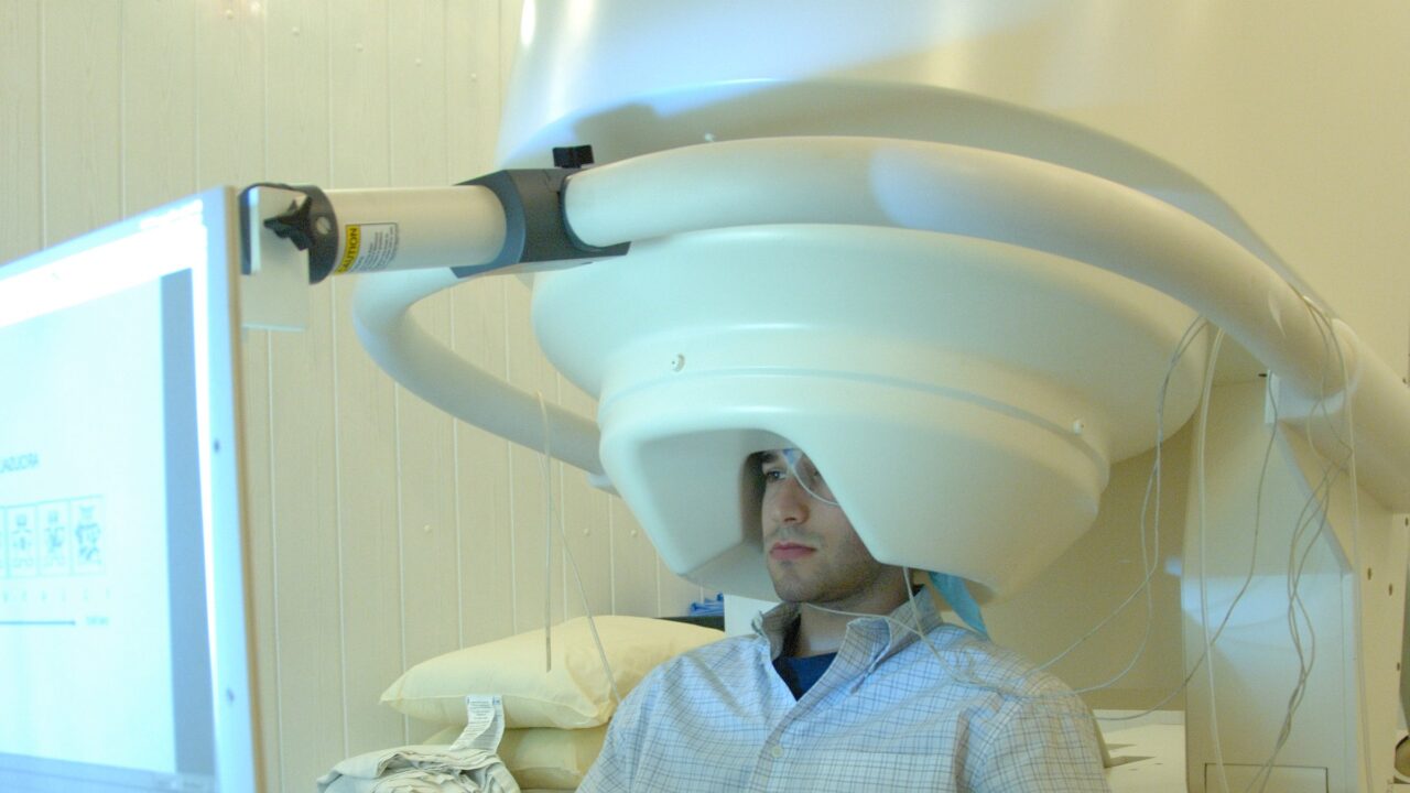
That finding comes from a magnetoencephalography study by Park, Correia, Ducorps and senior author Tallon-Baudry, published in Nature Neuroscience in 2014, volume 17, pages 612 to 618. Magnetoencephalography, usually shortened to MEG, records magnetic fields produced by cortical activity.
One point of wording matters here. The predictor was the heartbeat-evoked response, meaning the cortical activity time-locked to each beat, and not any property of the beat itself. Beat-to-beat variability of the heart rhythm, the electrocardiogram and peripheral blood pressure were all measured, and none of them separated the trials where people saw the grating from the trials where they did not.
The premise stated in the abstract is that bodily signals are constantly relayed up to the neocortex, so neural responses to bodily signals are likely to shape ongoing activity.
The task was a detection task run during MEG recording. A warning signal appeared on the screen, then the grating, which the abstract describes as faint, and then the volunteer delivered a response. The supplementary figure captions refer throughout to hits and misses, the trials where the grating was reported and the trials where it was not.
The authors' starting point, stated in the first line of the abstract, is that "Spontaneous fluctuations of ongoing neural activity substantially affect sensory and cognitive performance." Their added claim is that responses to bodily signals are part of those fluctuations.
So they compared the cortical response to heartbeats before stimulus onset, separately for hits and misses. The difference between the two fell in a 135 to 171 millisecond window in the heartbeat-locked signal. The captions show the data aligned to the T wave of the electrocardiogram.
The clearest numbers available outside the paywall sit in the fourth supplementary figure, which reports the analysis after cardiac artefact correction. Averaged across the cluster of sensors where the effect sat, the amplitude of the prestimulus heartbeat-evoked response differed between hits and misses across the 17 subjects (paired t-test, t16 = 4.81). The same figure compares both conditions against a separate resting session with the eyes open. The response was larger on hit trials than at rest (t16 = 3.46, p = 0.003) and smaller on miss trials than at rest (t16 = 2.39, p = 0.03).
The brain's reaction to a heartbeat was not simply bigger before a successful detection. It moved in both directions around an ordinary resting baseline.
On location, the abstract is specific. The authors write that "neural events locked to heartbeats before stimulus onset predict the detection of a faint visual grating in the posterior right inferior parietal lobule and the ventral anterior cingulate cortex, two regions that have multiple functional correlates and that belong to the same resting-state network."
A good deal of the work went into ruling things out, and the supplementary captions are where that shows. Interbeat interval variability, the beat-to-beat irregularity of the heart rhythm, decreased after the stimulus appeared, but it did not differ between hits and misses. The electrocardiogram itself, grand-averaged across subjects and locked to the T wave, showed no significant difference between the two. Respiration phase at the moment the stimulus appeared showed no preferred value in hits (p = 0.81) or in misses (p = 0.67). Blood pressure, computed over the same heartbeats used for the neural measure, showed no significant difference in systolic, diastolic, or the difference between systolic and diastolic pressure. Across a whole trial, blood pressure changed, but again not between seen and unseen.
The abstract compresses that into one line: "Neither fluctuations in measured bodily parameters nor overall cortical excitability could account for this finding."
Two further checks answer obvious objections. The first splits the group by heart rate. In 9 subjects with short interbeat intervals, averaging 730 milliseconds, and 8 with long ones, averaging 886 milliseconds, the hit versus miss difference held in both (t8 = 3.76, p = 0.006 and t7 = 3.19, p = 0.015). The caption draws the inference directly, noting that longer intervals "did not delay the effect, indicating that the effect corresponds to a differential response to the preceding heartbeat rather than a preparation of the next heartbeat."
The second is about artefact. The authors checked that the effect was not cardiac artefact, rerunning the analysis after removing cardiac components with independent component analysis, a technique that pulls the heartbeat's own signature out of the recording. The caption reports that the R and T waves could barely be seen in the corrected data, that the topography of the hit versus miss difference was not affected, and that the difference in the 135 to 171 millisecond window was preserved. Because the method extracts each participant's individual heartbeat shape, the caption argues, the effect does not depend on the morphology of any one person's beat.
The authors' reading is careful in its wording and large in its reach. "Neural events locked to heartbeats therefore shape visual conscious experience, potentially by contributing to the neural maps of the organism that might underlie subjectivity," they write. The abstract's final claim goes wider, that responses locked to a basic physiological input such as heartbeats underlie behaviourally relevant differential activation in cortical areas that do many different jobs.
Several readings the study does not support are easy to fall into. It does not show that a faster or stronger heartbeat improves vision, since interbeat interval variability, blood pressure and the electrocardiogram all failed to separate hits from misses. It does not place consciousness in the heart or in the body, since what differed between seen and unseen trials was cortical activity. And it does not establish that the heartbeat-evoked response caused detection. It predicted it, in this dataset, in these 17 people.
Seventeen is a small sample for a claim about conscious vision, and this is one experiment. The full text is behind a subscription, described on the journal's page as a preview of subscription content, so the account above rests on the abstract, the citation record and the supplementary figure captions that Nature publishes openly. Those supplementary analyses are detailed and the controls are serious. They are still controls on a single result rather than independent confirmations of it, and nothing in the openly available material describes an attempt to alter the heartbeat-evoked response and watch what happens to detection.
The paper's reference list does include a then-forthcoming piece by Park and Tallon-Baudry titled "The neural subjective frame: from bodily signals to perceptual consciousness," listed as in the press. The 2014 data themselves get as far as prediction. Moving the brain's response to a heartbeat and measuring what a person then sees would be a different experiment.
Study: Park HD, Correia S, Ducorps A, Tallon-Baudry C. "Spontaneous fluctuations in neural responses to heartbeats predict visual detection." Nature Neuroscience, volume 17, pages 612 to 618 (2014). DOI 10.1038/nn.3671.
Sources:
- Park HD, Correia S, Ducorps A, Tallon-Baudry C, "Spontaneous fluctuations in neural responses to heartbeats predict visual detection," Nature Neuroscience 17, 612 to 618, 2014, https://www.nature.com/articles/nn.3671
- Park HD, Correia S, Ducorps A, Tallon-Baudry C, same paper, citation record and supplementary figure captions, https://doi.org/10.1038/nn.3671