Movement and Reflexes Questions
1. A doctor strokes the bottom of a 35-year-old patient’s foot to elicit the plantar reflex. What would be a healthy response?
- A. The patient’s leg raises off of the table a few inches, then falls back down
- B. The patient’s toes curl downwards
- C. The patient’s big toe extends, and the other toes fan out
- D. The patient’s big toe extends, but the other toes curl downwards
Show Answer
Correct answer: B
A healthy plantar reflex involves the toes curling downwards. If the big toe extends and the other toes fan out, this is known as the Babinski sign. If it occurs in adults, it can be indicative of nervous system damage/dysfunction, often involving the corticospinal tract. The Babinski sign, however, is normal in infants. Some individuals may not respond to plantar stimulation; this is known as a "neutral" response. It doesn't necessarily indicate a problem, but may warrant further investigation.
2. A patient arrives at the hospital with symptoms of headache, dizziness, and an inability to move her right arm and leg. The doctor orders a CT scan; she suspects a stroke that has affected the patient’s left cerebral hemisphere. Why do you think the doctor assumed the left hemisphere was involved?
- A. Dizziness is more likely to occur with strokes that affect the left hemisphere
- B. The left hemisphere controls movement of the right side of the body
- C. Left hemisphere strokes tend to cause headaches, right hemisphere strokes do not
- D. Due to the brain's vasculature, most strokes affect the left hemisphere
Show Answer
Correct answer: B
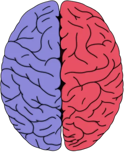
The doctor assumed the left cerebral hemisphere was involved due to the patient's inability to move her right arm and leg. Voluntary movement of the right side of the body is primarily controlled by signals that descend from the left cerebral hemisphere, and vice versa.
3. Damage to which of the following structures would be most likely to result in ataxia?
- A. amygdala
- B. hippocampus
- C. hypothalamus
- D. cerebellum
Show Answer
Correct answer: D
Ataxia involves impaired coordination of movement. It can have a variety of causes, but one region often implicated in ataxia is the cerebellum. The cerebellum is involved in many movement-related functions, including coordinating movement and maintaining balance. Thus, when there is cerebellar dysfunction (e.g., due to stroke), ataxia is a common result. For more info, see this article:
4. To test a patient’s corneal reflex, a doctor touches the patient’s right cornea with a piece of cotton. What response indicates a healthy corneal reflex?
- A. neither eye blinks
- B. only the patient's right eye blinks
- C. only the patient's left eye blinks
- D. both eyes blink
Show Answer
Correct answer: D
The corneal reflex is used clinically to test the integrity of the trigeminal and facial nerves and related structures, typically by touching the cornea with a piece of cotton. The healthy response to touching one cornea would be that both eyes would blink, but damage to the trigeminal or facial nerves or other related structures may cause a deficit in the reflex.
5. At the neuromuscular junction (where a neuron communicates with a muscle to cause muscle contraction), what is the neurotransmitter that is released to cause muscles to contract?
- A. dopamine
- B. glutamate
- C. serotonin
- D. acetylcholine
Show Answer
Correct answer: D
Neurons primarily release the neurotransmitter acetylcholine to cause contraction of skeletal muscle. Acetylcholine then binds to receptors in the muscle membrane to cause muscle contraction.
6. While you're walking across the floor, you step on something sharp. What reflex enables you to pull your leg away from the painful stimulus automatically, without having to consciously decide to do so?
- A. knee-jerk reflex
- B. plantar reflex
- C. triceps reflex
- D. withdrawal reflex
Show Answer
Correct answer: D
The withdrawal reflex (also known as the flexor withdrawal reflex) is an automatic, polysynaptic spinal reflex that causes a limb to quickly withdraw from a painful stimulus. Because the circuitry is contained within the spinal cord, the reflex does not require input from the brain and occurs without conscious control.
7. This neural pathway is the main pathway for voluntary movement. When you perform an action like picking up an object, neurons in this pathway carry signals from the motor cortex down to the spinal cord to cause muscle contractions. Which pathway is it?
- A. vestibulospinal tract
- B. spinothalamic tract
- C. dorsal columns-medial lemniscus pathway
- D. corticospinal tract
Show Answer
Correct answer: D
The corticospinal tract is the main pathway for voluntary movement.
8. Which of the following is a typical effect of lower motor neuron damage?
- A. spasticity
- B. over-responsive reflexes (hyperreflexia)
- C. weakness or paralysis
- D. increased muscle tone (hypertonia)
Show Answer
Correct answer: C
Lower motor neuron lesions often cause weakness or paralysis due to direct damage to the neurons that innervate skeletal muscle. This is frequently accompanied by decreased muscle tone and reduced or absent reflexes.
9. What is the significance of the neuromuscular junction?
- A. It is the site where sensory neurons transmit pain signals to the spinal cord
- B. It is the location where two motor neurons connect
- C. It is the area where nutrients are exchanged between blood and muscle
- D. It is the region where a motor neuron communicates with muscle to cause movement
Show Answer
Correct answer: D
The neuromuscular junction (NMJ) is a specialized synapse between a motor neuron and a skeletal muscle fiber. At the NMJ, acetylcholine released from the neuron binds to receptors on the muscle membrane, initiating an action potential that leads to muscle contraction.
10. Which basal ganglia pathway is primarily known for its role in facilitating voluntary movement?
- A. indirect pathway
- B. direct pathway
- C. corticospinal pathway
- D. vestibulospinal pathway
Show Answer
Correct answer: B
The direct pathway of the basal ganglia facilitates movement by reducing inhibition of the thalamus, which increases excitatory input to the motor cortex.
11. Which part of the cerebellum is MOST responsible for comparing actual limb position with the intended position during a movement?
- A. vestibulocerebellum
- B. spinocerebellum
- C. cerebrocerebellum
- D. dentate nucleus
Show Answer
Correct answer: B
The spinocerebellum receives information about limb position from the spinal cord and uses this information, for example, to compare where a limb is in space with where it should be if the movement were going as planned. If there is a discrepancy, the spinocerebellum can modify motor signals to correct any errors in the movement.
12. Which mechanism best explains how activation of the direct pathway of the basal ganglia is thought to facilitate movement?
- A. excitation of nuclei that inhibit thalamocortical activity
- B. direct excitation of motor cortex neurons by the striatum
- C. increased inhibitory signaling from the thalamus to the cortex
- D. inhibition of nuclei that inhibit thalamocortical neurons
Show Answer
Correct answer: D
The direct pathway promotes movement by inhibiting the globus pallidus internal and substantia nigra pars reticula, which normally inhibit the thalamus. This reduces inhibition of thalamocortical neurons, allowing increased cortical activity and facilitating movement.
13. The withdrawal reflex begins with these types of receptors:
- A. photoreceptors
- B. nociceptors
- C. baroreceptors
- D. chemoreceptors
Show Answer
Correct answer: B
Nociceptors are sensory receptors that detect painful or noxious stimuli. This is the critical first step in the withdrawal reflex, where a signal is sent from the nociceptors to the spinal cord, triggering a rapid, involuntary withdrawal of the body part from the source of the pain.
14. True or false: The main mechanism of the knee-jerk (patellar) reflex does NOT require input from the brain.
- A. True
- B. False
Show Answer
Correct answer: A
The knee-jerk reflex is a monosynaptic spinal reflex; the sensory neuron from the muscle spindle synapses directly onto the motor neuron in the spinal cord, causing contraction of the quadriceps. The brain is not required for the reflex itself, although it can modulate the strength of the response.
15. You step on a piece of sharp glass and reflexively pull your foot away from the painful stimulus. This reflex is known as the:
- A. knee-jerk reflex
- B. withdrawal reflex
- C. plantar reflex
- D. golgi tendon reflex
Show Answer
Correct answer: B
The withdrawal reflex is an automatic response enacted to withdraw a limb from a painful stimulus. If, for example, you were to touch a hot stove or step on a tack, the withdrawal reflex would cause you to pull your arm or leg away from the painful stimulus without having to consciously plan to do so. The reflex is also known as the flexor-withdrawal reflex.
16. During the knee-jerk (patellar) reflex, tapping the patellar tendon briefly stretches the quadriceps muscle. Which receptor detects this stretch and initiates the reflex arc?
- A. golgi tendon organ
- B. Pacinian corpuscle
- C. muscle spindle
- D. nociceptor
Show Answer
Correct answer: C
When the patellar tendon of the quadriceps muscle is stretched, the stretch is detected by stretch receptors known as muscle spindles that are found in the quadriceps muscle.
17. What enzyme is found at the neuromuscular junction and is responsible for breaking down acetylcholine?
- A. choline acetyltransferase
- B. monoamine oxidase
- C. catechol-O-methyltransferase
- D. acetylcholinesterase
Show Answer
Correct answer: D
The enzyme acetylcholinesterase is present at the neuromuscular junction; it breaks down acetylcholine and in the process terminates its effects on the muscle fiber, thus ending the communication between the motor neuron and the muscle fiber.
18. The knee-jerk (patellar) reflex involves contraction of the __________ muscle and inhibition of the _________ muscle.
- A. quadriceps; hamstring
- B. hamstring; quadriceps
- C. gastrocnemius; quadriceps
- D. quadriceps; gastrocnemius
Show Answer
Correct answer: A
The knee-jerk reflex causes immediate contraction of the quadriceps muscle to produce movement of the leg. Additionally, the sensory neurons traveling from muscle spindles stimulate interneurons that inhibit the activity of motor neurons that supply opposing muscles like the hamstring muscle. The stimulation of these inhibitory interneurons causes the action of the quadriceps muscle to be unopposed.
19. To test the corneal blink reflex, a doctor touches a patient's left eye with a wisp of cotton. When this is done, the patient's left eye blinks but the right eye does not. What is a likely explanation?
- A. there is damage to the patient's left trigeminal nerve
- B. there is damage to the patient's right trigeminal nerve
- C. there is damage to the patient's right facial nerve
- D. there is damage to the patient's left facial nerve
Show Answer
Correct answer: C
The corneal reflex involves sensory input from the ophthalmic branch of the trigeminal nerve (CN V) and motor output via the facial nerve (CN VII), which causes blinking. Since the left eye blinks, the sensory portion (left CN V) is functioning properly. The failure of the right eye to blink suggests that the right facial nerve (CN VII) is not carrying out the motor response.
20. A physician shines a light into a patient's right eye, and neither the right nor the left pupil constricts. When light is shined into the patient's left eye, both pupils constrict. Which of the following structures is most likely damaged?
- A. right optic nerve
- B. right oculomotor nerve
- C. left optic nerve
- D. left oculomotor nerve
Show Answer
Correct answer: A
The sensory limb of the pupillary light reflex is the optic nerve (CN II). If light shined into one eye fails to cause constriction in either eye, it suggests a problem with the sensory input—in this case, the right optic nerve. The motor (efferent) pathways are still intact, as the pupils can constrict when the other eye is stimulated.
21. Jean has a stroke that affects his right cerebellar hemisphere. Afterwards, he develops some movement and coordination problems. On which side of the body are these deficits likely to be more evident?
- A. right
- B. left
- C. there's no reason to think they'd be more evident on one than the other
Show Answer
Correct answer: A
While the right cerebral hemisphere primarily controls voluntary movement in the left side of the body, the right cerebellar hemisphere is primarily responsible for the right side of the body. Jean might experience symptoms such as impaired coordination, unrefined movements, tremors, etc., and they would likely be more prominent on the right side of the body.
22. Which of the following regions of the cerebellum is thought to be most directly involved with the maintenance of posture?
- A. dentate nucleus
- B. vermis
- C. cerebrocerebellum
- D. interposed nucleus
Show Answer
Correct answer: B
While various cerebellar regions are involved with posture, the vermis controls the trunk and axial muscles, making it the cerebellar region most directly responsible for maintaining posture and balance during standing and slow movement.
23. Where do most fibers of the corticospinal tract decussate (i.e., cross over)?
- A. anterior horn of the spinal cord
- B. medullary pyramids
- C. internal capsule
- D. cerebral peduncles
Show Answer
Correct answer: B
The corticospinal tract is a major pathway for carrying movement-related information from the brain to the spinal cord. Most corticospinal tract fibers decussate (i.e., cross over) at the level of the medullary pyramids in the lower brainstem, forming the lateral corticospinal tract.
24. Which of the following functions are the inferior olivary nuclei most closely associated with?
- A. Regulation of circadian rhythms
- B. Coordination and movement-related learning
- C. Control of emotional responses
- D. Maintenance of respiration
Show Answer
Correct answer: B
The inferior olivary nuclei play a key role in motor learning and coordination. They send climbing fiber inputs to the cerebellum, helping it fine-tune movements and adapt based on sensory feedback. This pathway is especially important for learning new motor skills and correcting errors during movement.
25. When motor signals are sent from the motor cortex to control movements of the head and face, which pathway do they primarily follow?
- A. Corticospinal tract
- B. Spinocerebellar tract
- C. Vestibulospinal tract
- D. Corticobulbar tract
Show Answer
Correct answer: D
The corticobulbar tract carries motor commands from the cerebral cortex to the motor nuclei of cranial nerves in the brainstem, which control muscles of the head and face. In essence, it does for the head and face what the corticospinal tract does for the rest of the body.
26. After a car accident, a patient suffers substantial damage to the right side of the cervical spinal cord. Her corticospinal tract fibers on the right are severely damaged. What motor deficit would you expect to see?
- A. weakness or paralysis on the right side of the body below the damage
- B. weakness or paralysis on the right side of the body above the damage
- C. weakness or paralysis on the left side of the body below the damage
- D. weakness or paralysis on the left side of the body above the damage
Show Answer
Correct answer: A
Damage to the right corticospinal tract in the spinal cord causes weakness or paralysis on the same side (ipsilateral) of the body below the lesion/damage.
27. Where do most of the fibers in the corticospinal tract decussate (i.e., cross to the other side of the nervous system)?
- A. in the medulla
- B. in the pons
- C. in the midbrain
- D. in the spinal cord
Show Answer
Correct answer: A
The upper motor neurons of the corticospinal tract leave the motor cortex and descend to the brainstem, continuing down into the medulla where the fibers form two bundles, known as the pyramids. At the base of the pyramids, about 90% of the fibers in the corticospinal tract decussate, or cross to the other side of the brainstem in a bundle called the pyramidal decussation.
28. Which of the following is a main input area of the basal ganglia?
- A. substantia nigra pars reticulata
- B. internal globus pallidus
- C. nucleus basalis of Meynert
- D. striatum
Show Answer
Correct answer: D
Much of the information the basal ganglia receive comes from the cerebral cortex and travels first to the striatum, which is a main input area of the basal ganglia. The main output nuclei of the basal ganglia are the internal globus pallidus and the substantia nigra pars reticulata, which send projections out of the basal ganglia to the thalamus as well as to nuclei in the brainstem.
29. Which of the following regions of the cerebellum is most involved in controlling posture?
- A. lateral hemisphere
- B. primary fissure
- C. superior cerebellar peduncle
- D. vermis
Show Answer
Correct answer: D
The vermis runs along the midline of the cerebellum, and is especially involved in controlling upright posture. It receives input from the vestibular system and spinal cord and helps to regulate the muscles of the trunk that maintain posture.
30. When a flashlight is shined into a patient’s right eye, neither pupil constricts. However, when the light is shined into the left eye, both pupils constrict normally. Based on these findings, which cranial nerve is most likely impaired?
- A. the right oculomotor nerve
- B. the left optic nerve
- C. the left oculomotor nerve
- D. the right optic nerve
Show Answer
Correct answer: D
If light is shined into the right eye and neither pupil constricts, it suggests a deficit in sensory input and may indicate damage to the right optic nerve.