How you see
- Lesson 1
- Lesson 2
- Lesson 3
- Lesson 4
- Lesson 5
- Lesson 6
- Lesson 7
- Lesson 8
- Lesson 9
- Lesson 10
- Lesson 11
- Lesson 12
Where you are
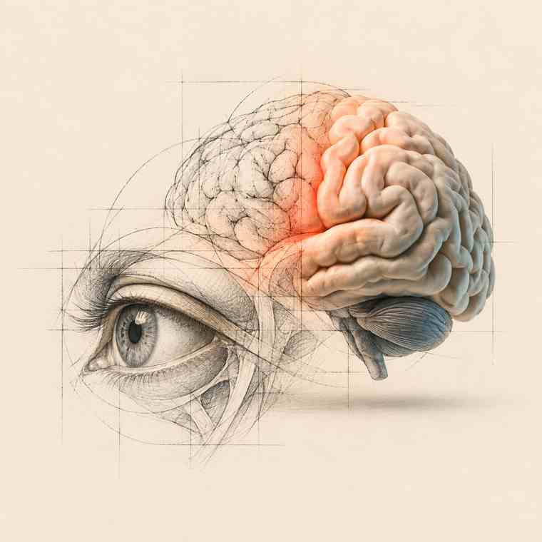
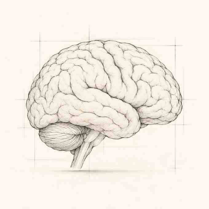
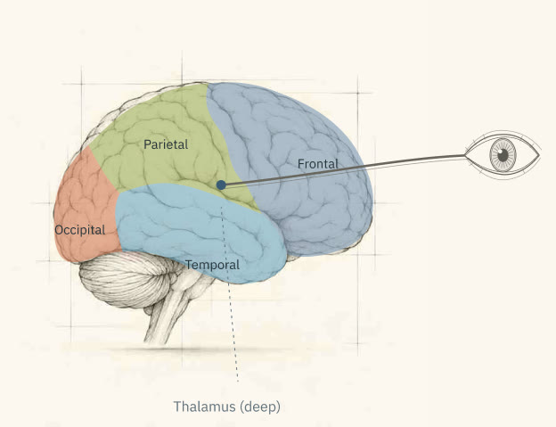
This is your brain, seen from the side.
It weighs about as much as a bag of sugar, has the texture of soft tofu, and runs on roughly the power of a dim light bulb. And right now, it's the thing reading this sentence. An organ studying itself is a strange situation. It's also the only way anyone has ever studied it.
On the map it's still blank, and that's deliberate. You won't memorize this map. You'll build it, one lesson at a time, starting from things you already do every day without thinking: seeing, getting scared, forgetting where you left your keys. By lesson 12, every region on this outline will mean something to you, because you'll have felt it at work.
We start with seeing. You're doing it right now, and it's where your brain has been quietly making things up your whole life.
The scene
It's 2 a.m. You get up for a glass of water, and in the doorway you freeze. Someone is standing in the corner of the room. You can make out the shoulders, the head tilted slightly forward. Your heart is already pounding.
You hit the switch. It's the coat you threw over the chair before bed.
Here's the strange part. For that half second, you didn't think someone might be there. You saw them. The shoulders were real to you. The light didn't reveal what had been in the corner all along. It swapped one picture for another, and your brain made both.
You might say: of course, it was dark. In good light, I see what's actually there.
You don't. Right now, in good light, there's a hole in your vision. One in each eye. Across a room, it's big enough to swallow someone's head. You've had it since you were born, and you've never noticed it, because your brain covers it over so well you'd never find it by accident.
Give me two minutes and I'll show you where it is.
Try it now
2 minutes
You need one eye, a screen and two minutes. The cross is where you look. The dot is what's going to disappear.
- Close your left eye.
- With your right eye, look straight at the cross and stay there. Don't move your eye to the dot, even when you're tempted. Just keep it in the corner of your vision.
- Slowly lean toward the screen, then back. Somewhere between 30 and 50 cm (12 to 20 inches)20 and 30 cm (8 to 12 inches), the dot will vanish.
Turn your phone sideways.
Turn your phone sideways.
It doesn't fade or blur. It's just gone, and the space where it was looks like plain background. Lean a little and it pops back. That's your blind spot.
Nothing happening? Check that your left eye is closed and that you're still looking at the cross. The pull to glance at the dot is strong, and the moment you look at it, it can't disappear.
Now the second test. Same rules: left eye closed, right eye on the cross. This time a line runs through the dot.
Turn your phone sideways.
Turn your phone sideways.
Find the distance where the dot vanishes, and notice what takes its place, still without moving your eye from the cross. Not a gap. Not a smudge. The line runs straight through, unbroken.
In that exact spot, the screen shows a dot, not a line. The line you saw there came from you.
Can't do this test?
Can't do this test?
With one eye closed and the other fixed on the cross, people can find a distance where the dot disappears and is replaced by the surrounding background. In the second test, the dot disappears again, but this time the line appears to continue through the space it occupied.
What just happened
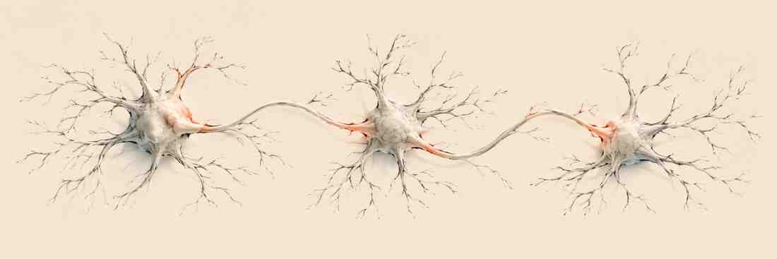
At the back of each eye, a cable plugs in: the optic nerve, about a million fibers carrying what the eye picks up to the brain. Where that cable leaves the eye, there's no room for light sensors. That patch is your blind spot. Everyone has one, in each eye.
Your brain doesn't leave the hole empty. It looks at what surrounds it and paints in whatever probably continues there: more line, more wall, more sky. With both eyes open, each eye also covers the other's gap.
And it goes well beyond that one spot. Hold your arm out and look at your thumbnail. A patch about that size is the only part of your view in sharp focus at any moment. Everything around it is blurry and poor in color. Your eyes jump several times a second, sampling one sharp patch after another, and your brain stitches them into the crisp, full-color scene you think you're simply looking at.
So the blind spot isn't an exception. It's just the one place where you can catch the stitching.
Where does the stitching happen? Follow the signal.
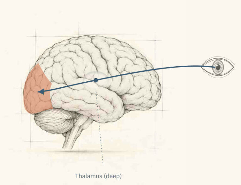
It leaves the eye through the optic nerve and travels to the very center of the brain, to a relay station called the thalamus. From there it's sent all the way to the back of your head, to the occipital lobe, where seeing begins: edges, lines, contrast, movement.
Put your hand on the back of your head. Roughly under your palm is where you're seeing this sentence, about as far from your eyes as the brain allows. Keep that in mind; the story below depends on it.
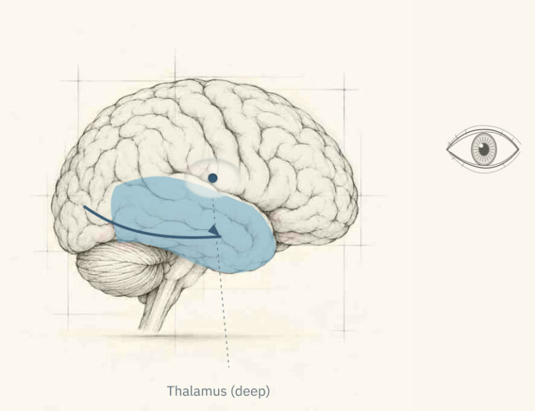
From there, the picture splits. One route runs down and forward into the temporal lobe, along the side of your head, above your ear. It answers what is it? This is where shapes become a face, a coat, a cup, your mother.
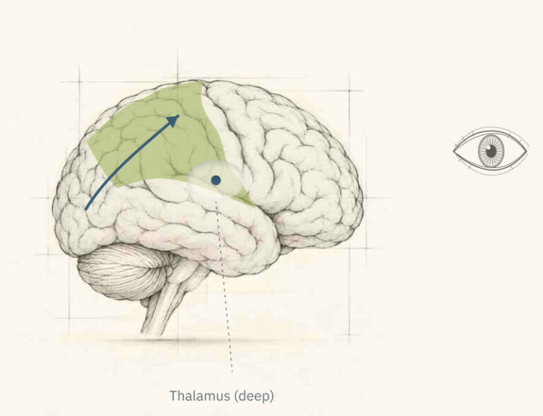
The other route runs up into the parietal lobe, at the top and toward the back. It answers where is it, and how do I reach it? It's why you can grab a cup without knocking it over.
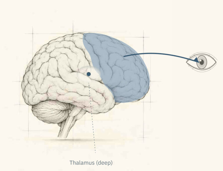
Remember those jumps your eyes make several times a second? Something chooses each one. That's the frontal lobe, behind your forehead, deciding about three times every second what deserves a closer look.
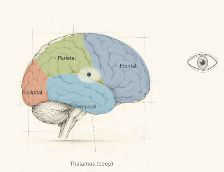
Those are the four lobes: occipital, temporal, parietal, frontal. They're regions of the cortex, the wrinkled outer layer of the brain, about as thick as two coins stacked. Seeing a single coat on a chair takes all four.
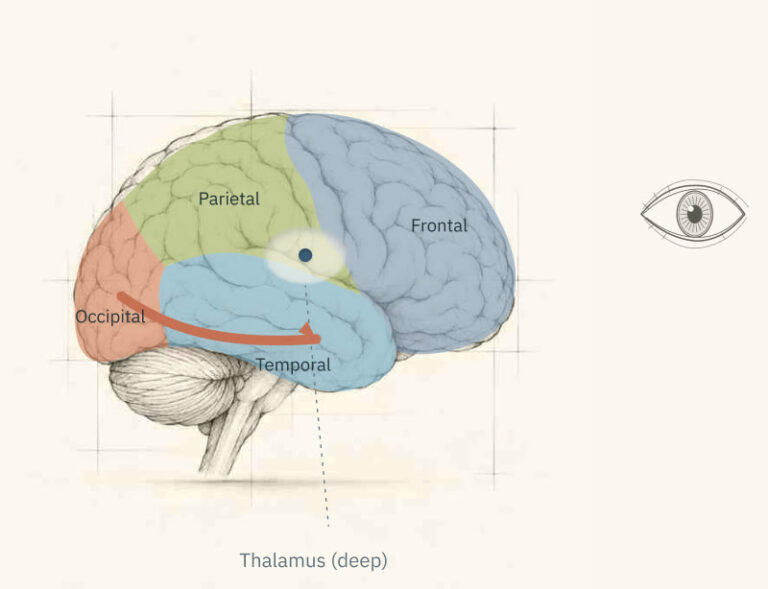
In the dark bedroom, the what route made its guess (a person) before there was enough light to be sure. Turning on the light gave it better information, and the guess changed. Your brain wasn't broken in that moment. It was doing what it always does, just with less to go on.

How it was discovered
The story: the doctor who drew the map from bullet wounds
During the Russo-Japanese War of 1904–1905, a Japanese eye doctor in his early twenties, Tatsuji Inouye, was handed a grim kind of paperwork. The army needed to know how much sight each wounded soldier had lost, to set his pension. So Inouye tested soldier after soldier, and among them were men who had been shot through the back of the head and survived.
They survived because of the weapon. The Russian army's Mosin-Nagant rifles fired small, very fast bullets that often went straight through the skull, leaving a clean hole going in and another coming out. In these men, the bullet had passed through roughly the spot where your hand was a few minutes ago. Each one had drawn a straight line through a soldier's brain.
Their eyes were fine. But part of the world was missing: a dark patch here, a blank quarter there. A hole in their vision, like the one you found, only bigger, and in a different place for each man.
Inouye worked it out like a surveyor. For each soldier, he located the two wounds and reconstructed the path the bullet had taken. Then he charted exactly which part of the man's vision had gone dark. Soldier after soldier, he matched the bullet’s path to the hole in the man’s vision.
The pattern was precise. Damage in one spot at the back of the brain always took away the same part of the visual world. Inouye had found a map: the scene in front of the eyes, laid out point by point across the back of the brain. And the map was lopsided. The center of vision took up far more space than the edges.
That same map is in your head right now. Its oversized center is the sharp patch you found with your thumbnail: small in your view, huge on the map. Your brain gives the most room to the part of the world you see in sharpest detail.
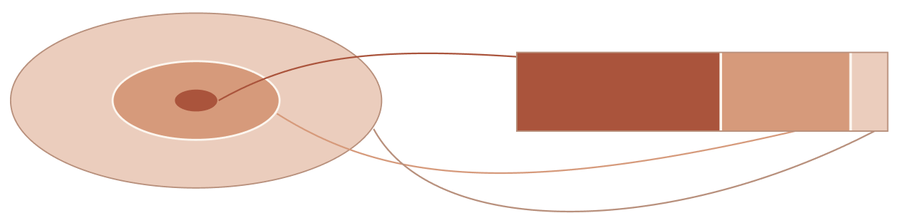
- Center
- Middle
- Outer
Schematic proportions · Center: within 10° of fixation. Horton & Hoyt, 1991; Horton, Economides & Adams, 2021.
Inouye published his findings in 1909. A few years later, during the First World War, the British neurologist Gordon Holmes, working independently, drew the same kind of map from soldiers wounded on the Western Front. His 1918 version stayed in use almost unchanged until 1991, when brain scans refined it.
What Inouye's soldiers revealed goes far beyond vision. Different parts of the brain do different jobs, so damage in one place tends to take away something specific and leave the rest. That's why the brain can be drawn as a map. It's the map you'll fill in over these twelve lessons, starting with the four lobes you've just met.
What you know now
Ten minutes ago, this outline was blank. Here's what's on it now.
1. Your eyes collect light; your brain builds what you see. It stitches small sharp patches into one clear scene and paints over the gaps. You caught it doing that at your blind spot.
2. Seeing travels. It starts at the back of your head, in the occipital lobe, then splits: down into the temporal lobe to work out what things are, up into the parietal lobe to work out where they are. Meanwhile the frontal lobe picks where your eyes go next.
3. In the brain, location matters. Different places do different jobs, which is how Inouye could match each soldier's wound to the hole in his sight. And some of those places hold maps of their own: the back of your head holds a map of your view, with the center taking up more than half of it.

Check yourself
Question 1 of 3
When the dot vanished in the experiment, why did you see an unbroken line instead of a gap?
Normally your other eye would cover the gap, but it was closed. And in that exact spot, the screen showed a dot, not a line. Your brain painted the line in.
Question 2 of 3
You're looking at a friend's face across a café. Which route recognizes that it's your friend?
The temporal route answers "what is it?" The parietal route answers "where is it?"
Question 3 of 3
One of Inouye's soldiers was shot through one specific spot at the back of the head. His eyes were unharmed. What happened to his sight?
Each spot at the back of the brain handles one part of what you see. That precision is how Inouye found the map, and it's the idea every following lesson builds on.
Go further
Want more on any stop along today's route? Most have an entry in Know Your Brain, our reference library. Each takes a few minutes and goes into the detail this lesson skipped. Read them now, or come back in a few lessons, when they'll make even more sense.
Reference
Reference
Reference
2-minute video