Know Your Brain: Basal Ganglia
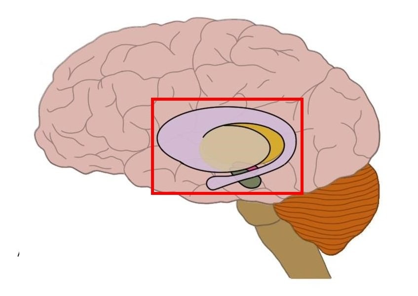
Where are the basal ganglia?

The basal ganglia are a group of structures found deep within the cerebral hemispheres. The structures generally included in the basal ganglia are the caudate nucleus, putamen, and globus pallidus in the cerebrum, the substantia nigra in the midbrain, and the subthalamic nucleus in the diencephalon.
The word basal refers to the fact that the basal ganglia are found near the base, or bottom, of the brain. The use of the word ganglia, however, is a bit of a misnomer according to contemporary neuroscience conventions. The term ganglion is used to describe a cluster of neurons, but it’s typically only used to refer to neurons in the peripheral nervous system (i.e., outside the brain and spinal cord). The word nucleus is generally used to describe clusters of neurons found in the central nervous system. Thus, the basal ganglia might more accurately be considered nuclei.
What are the basal ganglia and what do they do?
The separate nuclei of the basal ganglia all have extensive roles of their own in the brain, but they also are interconnected with one another to form a network that is thought to be involved in a variety of cognitive, emotional, and movement-related functions. The basal ganglia are best-known, however, for their role in movement.
The contributions of the basal ganglia to movement are complex and still not completely understood. In fact, the basal ganglia probably have multiple movement-related functions, ranging from choosing actions that are likely to lead to positive consequences to avoiding things that might be aversive. But the basal ganglia are most often linked to the initiation and execution of movements. One popular hypothesis suggests that the basal ganglia act to facilitate desired movements and inhibit unwanted and/or competing movements.
To understand how this might work, think about the action of reaching out to pick up a pencil. First, consider what’s happening in the moments before you extend your arm. Although it might seem like there would be very little movement-related activity going on in the brain at this point (because you are sitting still), your brain is actually constantly at work to inhibit unwanted movements (like jerking your hand involuntarily up in the air or suddenly turning your head to one side). The basal ganglia are hypothesized to play a critical role in this type of movement inhibition, as well as in the release of that inhibition when you do have a movement that you want to make (reaching for the pencil in this case).
After the movement begins, it’s also important that muscles that would counteract the desired movement remain relaxed. When you extend your arm to reach for the pencil, for example, you don’t want the muscles that flex your arm (to move it back towards your body) to be activated at the same time. The basal ganglia are thought to help to inhibit these types of contradictory movements, allowing for a reaching movement that’s smooth and fluid.
The intricacies of how basal ganglia activity leads to the facilitation of movement are still a bit unclear, but one popular hypothesis (which I’ll call the direct/indirect model for reasons that will be made clear below) suggests that there are different pathways in the basal ganglia that promote and inhibit movement, respectively. The direct/indirect model is centered around connections the basal ganglia (specifically the globus pallidus and substantia nigra) form with neurons in the thalamus. These thalamic neurons in turn project to the motor cortex (an area of the brain where many voluntary movements originate) and can stimulate movement via these connections. The basal ganglia, however, continuously inhibit the thalamic neurons, which stops them from communicating with the motor cortex—inhibiting movement in the process.
According to the direct/indirect model, when a movement is desired, a signal to initiate the movement is sent from the cortex to the basal ganglia, typically arriving at the caudate or putamen (which are referred to collectively as the striatum). Then, the signal follows a circuit in the basal ganglia known as the direct pathway, which leads to the silencing of neurons in the globus pallidus and substantia nigra. This frees the thalamus from the inhibitory effects of the basal ganglia and allows movement to occur. (For a more in-depth discussion of the direct and indirect pathways, see the next section below.)
There is also a circuit within the basal ganglia called the indirect pathway, which involves the subthalamic nucleus and leads to the increased suppression of unwanted movements. It is thought that a balance between activity in these two pathways may facilitate smooth movement.
Again, this is just one perspective on basal ganglia function, and despite the importance the basal ganglia are thought to have in movement, there is still much we need to learn to fully understand their contribution to it. We can see the importance of the basal ganglia to movement, however, when we look at cases where the basal ganglia have been damaged. In Parkinson's disease, for example, dopaminergic neurons of the substantia nigra degenerate. When this happens, the ability of the basal ganglia to cause the release of inhibition necessary to make a movement may be impaired. This may cause individuals with Parkinson's disease to have difficulty initiating movements, resulting in some of the symptoms associated with Parkinson's disease like rigidity and slow movement.
On the other hand, in a disorder like Huntington's disease, degeneration of basal ganglia circuits causes the inhibitory capabilities of the basal ganglia to be diminished. This may lead to the excessive activation of movement-related circuits, causing the jerky and writhing involuntary movements seen in Huntington's disease.
A balance between the ability to inhibit and facilitate movement is critical to making normal, smooth movements, and the proper functioning of the basal ganglia is essential to maintaining that balance. The basal ganglia, however, are also thought to have roles in habitual behavior, emotion, and cognition. Thus, in addition to movement disorders, the basal ganglia are also being investigated in attempts to understand disorders like Tourette's syndrome, schizophrenia, and obsessive-compulsive disorder.
More In-Depth Information
As mentioned above, the basal ganglia are often conceptualized as being organized into opposing pathways that facilitate and inhibit movement, respectively: the direct and indirect pathways. In this section, I will provide a more in-depth description of the direct and indirect pathways.
The Direct Pathway
The direct pathway of the basal ganglia is thought to regulate the activity of glutamate neurons in the thalamus that project to motor regions of the cerebral cortex. These neurons form excitatory connections with the motor cortex that are involved with the initiation of movement. When you want to make a movement, these thalamic neurons help to provide the excitation of the motor cortex that enables the movement to happen.
When you are not in the process of making a movement, however, your brain inhibits activity in these neurons---hypothetically to decrease the likelihood that you would end up making a movement that you didn't desire to make. For example, when you are sitting still reading this, it may seem like your brain doesn't have much to do when it comes to movement. But it is actually inhibiting a slew of undesired movements such as raising your arm into the air or turning your head away from what you're reading.
According to the classic model of basal ganglia function, one way this inhibition is achieved is due to the activity of neurons in the globus pallidus internal and substantia nigra pars reticulata, which project to the thalamus and maintain a steady release of the neurotransmitter GABA. GABA, which has inhibitory effects on neuronal activity, acts to suppress the activity of the thalamic neurons. This prevents them from communicating with the motor cortex, and inhibits movement.
When we want to make a movement, however, information about that movement is sent from the motor areas of the cerebral cortex to the striatum---the main input area of the basal ganglia. The pathway that extends from the cortex to the striatum is called the corticostriatal pathway. Glutamate neurons in this pathway excite neurons in the striatum, causing them to release GABA in the globus pallidus internal and substantia nigra pars reticulata. The released GABA inhibits the activity of these regions, and stops them from inhibiting the neurons in the thalamus that are involved with movement. This effectively opens a gate for movement to occur. Activity along this pathway tends to occur just prior to a movement, and thus has been linked to the facilitation of movement.
The substantia nigra pars compacta is thought to modulate the activity of the direct pathway. Neurons from the substantia nigra pars compacta travel to the striatum via the nigrostriatal pathway, and release dopamine in the striatum. One effect of this seems to be the facilitation of activity in the direct pathway.
The Indirect Pathway
The indirect pathway of the basal ganglia is hypothesized to play an opposing role to that of the direct pathway; thus, it is thought to be involved in the inhibition of movement. In other words, it is a circuit of the basal ganglia that acts to keep unwanted movements from occurring.
The indirect pathway involves GABA neurons that project from the external segment of the globus pallidus to a region called the subthalamic nucleus. These globus pallidus neurons typically exert an inhibitory effect on glutamate neurons in the subthalamic nucleus.
When the indirect pathway is activated by signals from the cerebral cortex, however, this causes the activation of GABA neurons in the striatum, which project to the globus pallidus external and inhibit the activity of neurons there. This keeps the globus pallidus external neurons from being able to inhibit neurons in the subthalamic nucleus.
The subthalamic nucleus neurons are activated by projections from the cerebral cortex, and they subsequently stimulate GABA neurons in the globus pallidus internal segment and substantia nigra pars reticulata. These GABA neurons in the globus pallidus internal and substantia nigra pars reticulata in turn project to the thalamus, inhibiting thalamic neurons that travel to motor regions of the cerebral cortex to stimulate movement. The inhibition of these thalamic neurons thus suppresses movement. This activity in the indirect pathway is thought to antagonize the activity of the direct pathway and act to keep unwanted movements from occurring.
Neurons from the substantia nigra pars compacta travel to the striatum via the nigrostriatal pathway, and they can modulate the activity of the indirect pathway through dopamine release in the striatum. One effect of this seems to be the inhibition of activity in the indirect pathway, which leads to the facilitation of movement. This is thought to be one reason why dopamine depletion in disorders such as Parkinson’s disease may lead to difficulties initiating movement. In Parkinson's disease, the degeneration of neurons in the substantia nigra leads to a reduction of activity in the globus pallidus external, and consequently increased activity in the subthalamic nucleus. This excessive subthalamic nucleus activity disrupts typical activity in the basal ganglia, possibly through excessive inhibition of neurons in the thalamus.
Modern perspectives
Originally, the basal ganglia were primarily considered to be part of the motor loop described above, which carries movement-related information from the cortex, to the basal ganglia, and back to the cortex. More recent hypotheses, however, suggest that there are multiple circuits that connect the basal ganglia with different areas of the cortex (not just motor regions). The diversity in the pathways of the basal ganglia through the cortex suggest the basal ganglia are involved in other functions besides movement, such as learning, attention, habit formation, motivation, and emotion. Thus, as mentioned above, the basal ganglia have a more expanded role than originally suggested, and their complete functions are still yet to be elucidated.
References:
Lanciego JL, Luquin N, Obeso JA. Functional neuroanatomy of the basal ganglia. Cold Spring Harb Perspect Med. 2012 Dec 1;2(12):a009621. doi: 10.1101/cshperspect.a009621. PMID: 23071379; PMCID: PMC3543080.
Purves D, Augustine GJ, Fitzpatrick D, Hall WC, Lamantia AS, McNamara JO, White LE. Neuroscience. 4th ed. Sunderland, MA. Sinauer Associates; 2008.
Learn more:


