Know Your Brain: Midbrain
Where is the midbrain?
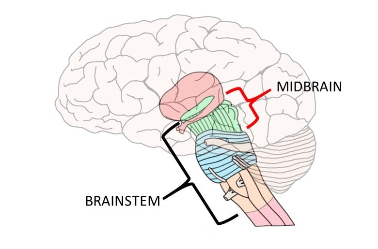
The midbrain is one of the three subdivisions of the brainstem; it is the most rostral of these subdivisions, or the one that is closest to the top of the brainstem. The midbrain connects the brainstem to the diencephalon at a location sometimes called the midbrain-diencephalon junction.
What is the midbrain and what does it do?
Although it is a relatively small section of neural tissue, the midbrain contains a long list of nuclei, tracts, nerves, and other structures---each with its own diverse catalog of functions. Thus, any attempt to define all of the actions of the midbrain in just a few sentences, paragraphs, or even pages is inherently inadequate. Rather than attempt to do that, I will simply discuss some of the most prominent anatomical features of the midbrain and some of their putative functions.
One of the most noticeable external features of the midbrain is the presence of four bumps on its posterior surface (the side that faces the back of the brain). Those bumps are indicative of the presence of four large underlying clusters of neurons; the upper pair of those clusters are known as the superior colliculi and the lower pair are known as the inferior colliculi. The superior colliculi are thought to be involved in directing behavioral responses toward stimuli in the environment, while the inferior colliculi are best known for their role in auditory processing.
The anterior surface of the midbrain is marked by the presence of the crura cerebri (plural for crus cerebri), two large bundles of axons that travel along the base of the midbrain as they stretch from the pons to the cerebral hemispheres. They contain fibers that are part of important motor pathways like the corticospinal and corticobulbar tracts. The crura cerebri are sometimes called the cerebral peduncles, although this term is generally used to refer to a larger area that includes the crura cerebri as well as much of the rest of the midbrain.
The midbrain is often divided into three regions. At the level of the midbrain, the fourth ventricle has narrowed to form the cerebral aqueduct, a channel that connects the fourth ventricle with the third ventricle. The region of the midbrain posterior to the cerebral aqueduct is called the tectum, which means "roof" in Latin. The tectum consists primarily of the superior and inferior colliculi.
The area of the midbrain anterior to, or in front of, the cerebral aqueduct is called the tegmentum. The tegmentum contains a variety of ascending and descending tracts that pass through the midbrain, such as the medial lemniscus and the anterolateral tracts. Fibers from the superior cerebellar peduncles, the major efferent pathway from the cerebellum, decussate in the midbrain. Some of these fibers project to a midbrain nucleus called the red nucleus, which is thought to play an important role in motor coordination.
There are several other important nuclei and neuronal clusters in the midbrain tegmentum. For example, the midbrain tegmentum contains nuclei for two cranial nerves: cranial nerve III (oculomotor nerve) and cranial nerve IV (trochlear nerve). It also contains neurons that are part of the raphe nuclei---clusters of serotonin-producing neurons found in the brainstem that send serotonin throughout the central nervous system. Additionally, one of the largest collections of dopamine-producing neurons in the brain, the ventral tegmental area, is located in the midbrain tegmentum.
The third region of the midbrain is made up of two structures called the basis pedunculi. They cover the anterolateral portions (to the front and toward the sides) of the brainstem. The basis pedunculi include the crura cerebri (discussed above) as well as the substantia nigrae, which---like the ventral tegmental area---contain large collections of dopamine-producing neurons.
Finally, the area surrounding the cerebral aqueduct is called the periaqueductal gray, or PAG. The PAG has long been recognized for its role in pain inhibition, although it is also thought to be involved in many other functions ranging from emotional responses to the production of vocalizations.
References:
Haines DE. Fundamental Neuroscience for Basic and Clinical Applications. 4th ed. Philadelphia, PA: Elsevier; 2013.
Vanderah TW, Gould DJ. Nolte's The Human Brain. 7th ed. Philadelphia, PA: Elsevier; 2016.