Associating brain structure with function and the bias of more = better
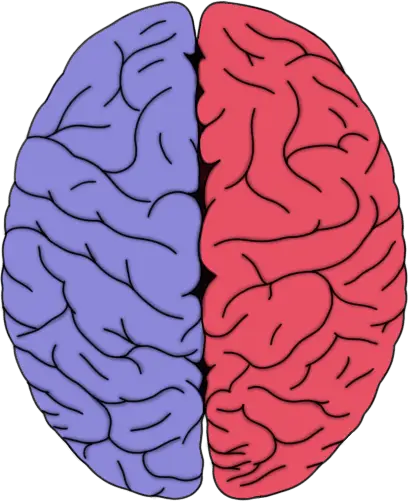
It seems that, of all of the behavioral neuroscience findings that make their way into popular press coverage, those that involve structural changes to the brain are most likely to pique the interest of the public. Perhaps this is because we have a tendency to think of brain function as something that is flexible and constantly changing, and thus alterations in function do not seem as dramatic as alterations in structure, which give the impression of being more permanent.
After all, until relatively recently it was believed that we are born with a fixed number of neurons--and that was it. From the end of neural development through the rest of one's life it was thought that no new neurons were produced; thus, the inevitable occurrence of neuronal death ticked off a slow but inexorable decline into cognitive obscurity that we were helpless to prevent. We now know that this is not true, however, and that there are new neurons produced throughout one's lifespan in certain areas of the brain.
Regardless, perhaps that outdated thinking on the immutability of the brain causes people to be especially impressed by the mention of some activity changing the structure of the brain, because there is no shortage of articles in the popular press covering studies that involve changes to brain structure. Just since the beginning of 2015, there have been major news stories about methamphetamine, smoking, childhood neglect, and head trauma in professional fighters all leading to reductions in grey matter, as well a more positive story that music training in children can lead to increased grey matter in some areas of the cortex. And it seems like stories about meditation's ability to increase grey matter are constantly being recycled among blogs and popular news sites. In all of these stories it is either implied or stated without much supporting evidence that adding more grey matter to a part of the brain increases cognitive function, while losing grey matter decreases it. For example, in a story about smoking and its effects on the cortex, the reduction in grey matter was described in this way: "As the brain deteriorates, the neurons that once resided in each dying layer inevitably get subtracted from the overall total, impairing the organ’s function."
Of course, in neurodegenerative diseases like Alzheimer's disease, we know that loss of brain tissue corresponds to increasingly more severe deficits. But what do we know for sure about non-pathological changes in the structure/size of the brain, and how they are associated with function? Is it widely accepted that more grey matter is equivalent to improved function and less to diminished function? Contrary to what you might conclude if you read some recent news articles--and in many cases, the studies they summarize--on these subjects, we have not found a consistent formula for predicting how changes in structure will affect function. And so, it is not a universal rule that more is better when it comes to grey matter.
More brain mass = better brain function?
The hypothesis that larger brain size is associated with increased mental ability can be traced back at least to the ancient Greeks (possibly to the physician Erasistratus). It has had the support of a large share of the scientific community since the 1800s when some of the first formal experiments into the matter were conducted by Paul Broca. Broca recorded the weights of autopsied brains and found a positive association between education level and brain size. These findings would later be cited by Charles Darwin as he made a case for the large brain of humans as an evolved trait responsible for our superior intelligence compared to other species.
Indeed, the hominid brain has had a unique evolutionary trajectory. It is thought to have tripled in size about 1.5 million years ago, in the process creating a large disparity between our brains and those of our non-human primate relatives like the great apes. This rapid brain expansion, along with our highly developed cognitive abilities, has led many to suggest our brain growth was directly connected to increased capacities for intellect, language, and innovation. Humans don't have the biggest brains in the animal kingdom, however (that distinction belongs to sperm whales, which have brains that weigh about six times what ours do), but it is thought that the way the human brain grew--by adding more to cortical areas that are devoted to conceptual, rather than perceptual and motor processing--may have been responsible for our accelerated gains in intellectual ability.
Thus, while brain size has come to be considered an important indicator of the intelligence of humans in comparison to other species, it is thought that the cerebral cortex is really the defining feature of the human brain. The cortex makes up about 80% of our brain, a proportion that is much higher than that seen in many other mammalian species. The intricate folding and complex circuitry of the cerebral cortex may contribute to the greater intellectual capabilities we have compared to other species.
The advent of neuroimaging and its refinement over the last 15 years have allowed us to test in vivo the hypothesis that brain size among individuals corresponds to intelligence and, more specifically, that the thickness of the cerebral cortex is especially linked to cognitive abilities. Many of the results have supported these hypotheses. For example, a recent analysis of 28 neuroimaging studies found a significant average correlation of .40 between brain size and general mental ability. Another study of 6-18 year-olds in the United States found cortical thickness in several areas of the brain to be associated with higher scores on the Wechsler Abbreviated Scale of Intelligence. Choi et al. (2008) also found a positive correlation between cortical thickness and intelligence measures like Spearman's g; in addition they were able to explain 50% of the variance in IQ scores using estimates of cortical thickness in conjunction with functional magnetic resonance imaging data.
Recent studies have also allowed us to observe that brain size is not completely static, and that experience (as well as age) can alter the size or structure of certain parts of the brain. For example, one well-known study looked at the brains of London taxi drivers and found that taxi drivers had larger hippocampi compared to control subjects (hypothetically because the hippocampus is involved in spatial memory). The size of hippocampi was also correlated with time spent driving a taxi, suggesting it may have been the experience of navigating a cab through London that promoted the hippocampal changes, and not just that people with better spatial memory were more likely to become taxi drivers. In another study, participants underwent a brain scan, then spent a few months learning how to juggle, then had a second brain scan. The second scan showed increased grey matter in areas of the cortex associated with perceiving visual motion. When they stopped juggling, then had a third brain scan a few months later, the size of these areas had again decreased.
Based on such results, a number of studies have also looked at how certain activities might affect cortical thickness, with the assumption that increased cortical thickness represents an enhancement of ability. For example, an influential study published in 2005 by Lazar et al. examined the brains of 20 regular meditators (with an average of about 9 years of meditation experience) and compared the thickness of their cortices to that of people with no meditation experience. The investigators found that the meditators had increased grey matter in areas like the prefrontal cortex and insula; the authors interpreted this as being indicative of enhanced attentional abilities and a reduction of the effects of aging on the cortex, among other things.
Problems with the increased mass = improved function hypothesis
But there are a few problems with conclusions like those made in the meditation study conducted by Lazar et al. The first is that in the Lazar et al. study--and in many studies that look at brain structure and associate it with function--brain structure was only assessed at one point in time. The issue with only looking at brain structure once is that it doesn't allow one to determine if the structure changed before or after the experience. In other words, in the Lazar et al. study perhaps increased cortical thickness in prefrontal areas was associated with personality traits that made individuals more likely to enjoy meditating, instead of meditating causing the structure of the cerebral cortex to change.
Regardless, even if we knew the behavior (e.g. meditation) came before the structural changes, it still would be unclear how the structural modifications translated into changes in function. Postulating, as Lazar et al.did, that the changes may have been associated with an increased ability to sustain attention or be self-aware is a clear example of confirmation bias, as the authors are interpreting results in such a way as to support their hypothesis when truly they lack the evidence to do so. However, even if we agreed with the specious reasoning of Lazar et al. that "the right hemisphere is essential for sustaining attention" and thus structural changes to it are especially likely to affect attention, it is far from conclusive that increased cortical thickness in general represents increased function.
In fact, there are myriad examples in the literature that suggest there is not always a positive correlation between cortical thickness and cognitive ability. For example, a study involving patients with untreated major depressive disorder found they had increased cortical thickness in several areas of the brain. Another study detected increased cortical thickness in frontal, temporal, and parietal lobes in children and adolescents born with fetal alcohol spectrum disorder. A 2011 investigation involving binge drinkers observed thicker cortices in female binge drinkers, and thinner cortices in male binge drinkers. And a report examining the effects of education level on cortical thickness found those with higher education levels actually had thinner cortices. This is just a small sampling of the many studies indexed on PubMED that do not support the increased mass = increased function hypothesis. The reasons for this lack of consistent support could be numerous, ranging from confounds to measurement problems. But the important point is that there is no axiom that increased grey matter will result in improved functionality.
Of course this doesn't mean there is never an association between brain volume or cortical thickness with brain function. We should, however, be interpreting studies that identify such associations with caution. And we should be even more wary of popular press articles that summarize such studies, for when these studies are reported on by the media, the complexity of the association between structure and function--as well as the cross-sectional limitations of many of the studies--are rarely taken into consideration.
There are some methodological approaches to these types of studies that would allow investigators to improve their ability to make confident interpretations of the results. First, an emphasis should be placed on using longitudinal designs. In other words, an initial brain scan should be conducted first to get a baseline measure of brain structure. Then the activity (e.g. meditation) should be performed for a period of time before brain structure is assessed at least one more time. This reduces the weight that must be given to the concern that changes in structure predated changes in function. Additionally, it is best for some measure of function to be taken along with an assessment of brain structure to support any suggestion that the structural changes correspond to a functional change.
Many studies are already starting to utilize such approaches. For example, a recent study by Santarnecchi et al. examined the effects of mindfulness meditation on brain structure. The researchers conducted brain scans before and after an 8-week Mindfulness Based Stress Reduction (MBSR) program, using participants who had never meditated before. Participants also underwent psychological evaluations before and after the program. The participants experienced a reduction in anxiety, depression, and worry over the course of the MBSR course. But they also showed an increase in cortical thickness--in the right insula and somatosensory cortex. The Lazar et al. study noted an increase in volume of the right insula as well, suggesting there may be something to this finding. And because of the longitudinal design of the study by Santarnecchi et al., along with the fact that the participants were not regular meditators, we can have more confidence that the structural changes seen came as a result of--instead of predated--the meditation practice.
As can be seen from this example where, despite the methodological problems of the Lazar et al. study, some of the results were later supported, a too liberal interpretation of such results does not suggest the findings are baseless. If we are going to have confidence in the value of associations between structure and function, however, more emphasis must be placed on using study designs that allow one to infer causality. And, when it comes to the interpretation of results, a conservative approach should be advocated.
Lazar, S., Kerr, C., Wasserman, R., Gray, J., Greve, D., Treadway, M., McGarvey, M., Quinn, B., Dusek, J., Benson, H., Rauch, S., Moore, C., & Fischl, B. (2005). Meditation experience is associated with increased cortical thickness NeuroReport, 16 (17), 1893-1897 DOI: 10.1097/01.wnr.0000186598.66243.19

