Know Your Brain: Retina
Where is the retina?
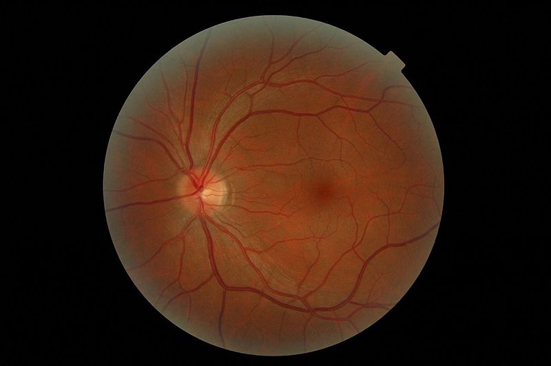
The retina is a collection of cells at the back of the eye where the processing of visual information begins. It is only about as thick as a razor blade, but contains several layers of cells that are made up of a number of different cell types. The retina forms from brain tissue during development, and is considered part of the central nervous system.
What is the retina and what does it do?
When light reaches the back of the eye, it then enters the cellular layers of the retina. The design of the retina is somewhat counter-intuitive as the cells that detect and respond to light, known as photoreceptors, are located at the very back of the retina. This means that light must travel through several layers of cells before reaching the point where visual processing begins.
There are two types of photoreceptors: rods and cones. Rods have poor resolution, but are very sensitive to light. They allow us to see in dim light, but don't allow for the perception of color. Cones, on the other hand, have high resolution but are not as sensitive; they allow us to perceive color under normal lighting conditions. Throughout most of the retina, rods outnumber cones. In one area called the fovea, however, the number of cones is increased 200x, and there are little to no rods. The fovea represents the area of the retina that provides our highest acuity vision, and thus is at the center of our gaze.
When light hits photoreceptors, it interacts with a molecule known as a photopigment, which responds by undergoing a chemical change. In this case the chemical change begins a chain of events that serves to propagate the visual signal. Activation of the photopigment leads to a change in membrane potential and a modification in the amount of neurotransmitter released from photoreceptor cells. This process of translating visual information into a signal that can be passed through the nervous system is called phototransduction.
Phototransduction leads to a signal being transmitted to cells called bipolar cells, which connect photoreceptors to ganglion cells. Bipolar cells send the signal to ganglion cells, which travel out of the eye in a large cluster through an opening called the optic disc. After leaving the retina, the ganglion cell fibers are called the optic nerve. The optic nerve carries visual information toward the brain to be processed.
The optic disc doesn't contain any photoreceptors, and so represents an area on the retina that can't process visual information, creating a natural blind spot. The blind spot is actually relatively large; although the size varies from individual to individual, it can be upwards of 2mm in diameter and cover an area of your visual field equivalent to the width of your four fingers when held at arm's length. However, we usually don't ever notice our blind spot. The brain uses information from surrounding photoreceptors and the other eye to fill in the gaps in the images that are processed by the retina.
If you want proof that the blind spot is there, take a look at the X and O below. Keep your computer screen at about arm's length and cover your left eye. Keep your right eye on the O, then slowly move your face towards the screen, all the while continuing to look only at the O. At some point, the image of the X in your peripheral view should disappear. This is because at that point, the image of the X is falling on the blind spot of your right eye.
O....................................................................................X
There are two other cell types in the retina that should be mentioned: horizontal and amacrine cells. Horizontal cells receive input from multiple photoreceptor cells. They use that input to integrate signaling from different populations of photoreceptor cells, make adjustments to the signals that will be sent to bipolar cells, and regulate activity in photoreceptor cells themselves. Amacrine cells receive signals from bipolar cells and are involved in the regulation and integration of activity in bipolar and ganglion cells.
Reference (in addition to linked text above):
Nolte J. The Human Brain: An Introduction to its Functional Anatomy. 6th ed. Philadelphia, PA. Elsevier; 2009.