Know Your Brain: Amygdala
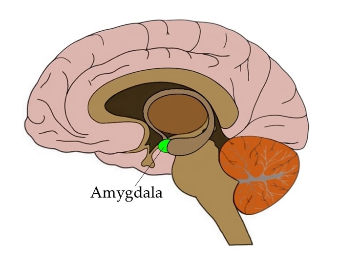
Where is the amygdala?

The amygdala is a collection of nuclei found deep within the temporal lobe. The term amygdala comes from Latin and translates to "almond," because one of the most prominent nuclei of the amygdala has an almond-like shape. Although we often refer to it in the singular, there are two amygdalae—one in each cerebral hemisphere.
What is the amygdala and what does it do?
The amygdala is recognized as a component of a group of brain structures referred to collectively as the limbic system, and is thought to play important roles in emotion and behavior. It is best known for its role in the processing of fear, although as we’ll see, this is an oversimplified perspective on amygdala function.
Our modern understanding of amygdala function can be traced back to the 1930s, when Heinrich Kluver and Paul Bucy removed the amygdalae of rhesus monkeys and saw drastic effects on behavior. Among other things, the monkeys became more docile and seemed to display little fear. The constellation of behavior that resulted from amygdalae removal was called Kluver-Bucy syndrome, and it led to the amygdala being investigated for its role in fear.
Since, the amygdala has become best known for its role in fear processing. When we are exposed to a fearful stimulus, information about that stimulus is immediately sent to the amygdala, which can then send signals to areas of the brain like the hypothalamus to trigger a "fight-or-flight" response (e.g., increased heart rate and respiration to prepare for action).
Interestingly, research suggests that information about potentially frightening things in the environment can reach the amygdala before we are even consciously aware that there’s anything to be afraid of. There is a pathway that runs from the thalamus to the amygdala, and sensory information about fearful stimuli may be sent along this pathway to the amygdala before it is consciously processed by the cerebral cortex. This allows for the initiation of a fear reaction before we even have time to think about what it is that’s so frightening.
This type of reflexive response can be useful if we really are in great danger. For example, if you are walking through the grass and a snake darts out at you, you don't want to have to spend a lot of time cognitively assessing the danger the snake might pose. Instead, you want your body to experience immediate fear and jump backward without having to consciously initiate this action. The direct pathway from the thalamus to the amygdala may be one way to achieve this type of response.
In addition to its involvement in the initiation of a fear response, the amygdala also seems to be very important in forming memories that are associated with fear-inducing events. For example, if you take mice with intact amygdalae and play a tone right before you give them an uncomfortable foot shock, they will very quickly begin to associate the tone with the unpleasant shock. Thus, they will display a fear reaction (e.g., freezing in place) as soon as the tone is played, but before the shock is initiated. If you attempt this experiment in mice with lesions to the amygdalae, however, they display an impaired ability to "remember" that the tone preceded the foot shock. You can play the tone and they will continue about their business as if they have no bad memories associated with the noise.
It shouldn't be too surprising (given its role in fear processing) that the amygdala might also play a role in anxiety. While fear is considered a response to a threat that is present, anxiety involves the dread that accompanies thinking about a potential threat—one that may or may not ever materialize. A number of studies suggest that the amygdala is involved in experiencing anxiety, and that it may be overactive in people with anxiety disorders. However, as is the case with most human behaviors, anxiety likely involves a network of brain areas, so activity in the amygdala doesn’t tell us all we need to know about the emotion.
Although the amygdala is well-known for its role in fear responses, there is now a great deal of evidence that suggests its contribution to behavior is much more complex. For example, the amygdala seems to be involved with the formation of positive memories, like earning a reward in an experiment. And damage to the amygdala can impair the ability to form these positive memories, just like it can affect the ability to form memories about negative events like the foot shock mentioned above.
Because of research like this, researchers have been forced to expand the role of the amygdala beyond that of just a threat detector/fear generator. One popular perspective suggests that the amygdala is involved with evaluating things in the environment to determine their importance—whether their value is positive or negative—and generating emotional responses to those stimuli that are considered important. It also may be involved in the consolidation of memories that have some strong emotional component, regardless of whether the associated emotions are pleasant or unpleasant. Thus, our understanding of the function of the amygdala is still evolving, and we likely have much more to learn before we can fully catalog the activities of this complex structure.
More In-Depth Information
History
The experiments of Heinrich Kluver and Paul Bucy in the 1930s eventually led to the amygdala being identified as an area of interest in the neuroscience of human behavior. Kluver had been studying (and taking) the psychedelic drug mescaline, and was interested in what part of the brain might be responsible for producing the unique hallucinatory effects of the drug. Kluver hypothesized that the brain region in question might reside in the temporal lobe, because large doses of mescaline given to monkeys could produce side effects that resembled the symptoms of a type of epilepsy called temporal lobe epilepsy.
Kluver recruited a young neurosurgeon named Paul Bucy to help him explore his hypothesis by surgically removing parts of the temporal lobe in monkeys. If the temporal lobe was critical for the effects of mescaline, Kluver hypothesized, then removing enough brain tissue from the region should render the drug ineffective.
The first monkey Kluver and Bucy operated on was an aggressive and unruly monkey named Aurora. Bucy removed most of Aurora's right and left temporal lobes, and afterwards he and Kluver noted some drastic changes to Aurora's personality. Previously, Aurora had been hostile and difficult to control, but now she was placid and easy to work with. In fact, she seemed almost incapable of anger; she also displayed no fear reactions. When Kluver and Bucy published a description of Aurora's changes in personality, it was the first well-known study to link the temporal lobe to emotion. The constellation of behavior that appeared after temporal lobe damage came to be known as Kluver-Bucy syndrome.
A couple of decades later, another scientist named Larry Weiskrantz found that he could elicit Kluver-Bucy syndrome in monkeys just by removing the amgdalae (which are found in the temporal lobes). Weiskrantz's work led other researchers to focus more on the amygdala's role in emotional responses.
While Weiskrantz hypothesized that the amygdala might be involved in enabling monkeys to respond emotionally to both positive and negative stimuli in the environment, many researchers after him focused more on the negative emotions linked to the structure. A large number of studies specifically investigated the amygdala's role in fear.
Often these studies involved what is known as a fear conditioning paradigm. Fear conditioning experiments promote the learning of a fearful response to something that previously didn't inspire any fear. These experiments typically involve taking a subject (such as a rodent) and exposing it to a stimulus (such as a beeping tone) that the animal has no positive or negative experience with. Next, the neutral stimulus is paired with something the animal will definitely perceive as negative (such as a mild electrical shock). If this is done enough times, the rodent will eventually begin to display signs of fear when the tone is played, even when it isn't immediately followed by the shock.
Researchers found that damage to the amygdala disrupts fear conditioning. In other words, if you damage a rat's amygdalae and then put it into a fear conditioning experiment, it won't learn to fear the tone—no matter how many times the tone is paired with a shock. Further research in rodents found that neurons in the amygdala are highly active when rodents hear a tone that has been linked to a fear-inducing stimulus. And studies in humans helped to confirm the role of the amygdala in learning about and experiencing fear-inducing things.
As mentioned above, subsequent research has demonstrated that the amygdala is involved in much more than just fear. For example, more recent experiments have shown that the amygdala also plays a role in learning about positive things (like rewards), and amygdala damage can disrupt the ability to form memories about those positive stimuli. Neuroscientists today generally support a perspective that the amygdala is important in creating emotional responses to both positive and negative things in our environment, as well as in forming memories about those emotionally-salient things.
Disorders involving the amygdala
There are several neurological disorders associated with damage to the amygdala. One, as discussed above, is Kluver-Bucy syndrome. Kluver-Bucy syndrome is rare in humans, but can occur after brain trauma, neurodegenerative disease, or an infection that reaches the brain. The symptoms vary from case to case, but might include placidity, an irresistible urge to put various objects (appropriate and inappropriate) in the mouth (also known as hyperorality), and an uncontrollable appetite.
Urbach-Wiethe disease is a rare genetic disorder that can cause calcification of brain tissue in the temporal lobes; this calcification can cause damage to the amygdalae. While Urbach-Wiethe disease is an exceedingly rare condition, it is thought to be the cause of amygdala damage in one of the best-known medical cases alive today: SM. SM, who is only known by her initials to protect her anonymity, has a well-documented inability to experience fear. Over the past several decades, researchers have put SM into a variety of experimental conditions designed to elicit fear. Only one—forcing her to breathe air that was about 35% carbon dioxide (a solution that causes people to struggle to breathe and often elicits panicked reactions)—led to a fearful reaction from SM. SM has Urbach-Wiethe disease, and it has caused severe damage to her amygdalae. Because of her inability to experience most types of fear coupled with her amygdala damage, SM is commonly used as an demonstration of the important role the amygdala plays in fear.
The amygdala is also thought to be involved in certain types of temporal lobe epilepsy, which might explain some characteristics of temporal lobe seizures, such as feelings of fear and strong emotional memories. Additionally, the amygdala is implicated in some of the cognitive and behavioral symptoms of neurodegenerative dementias, like Alzheimer's disease. Studies suggest, for example, that the death of neurons in the amygdala in Alzheimer's disease may be a substantial contributor to the memory loss characteristic of the condition.
A long list of studies have suggested the involvement of the amygdala in various psychiatric disorders. For example, as mentioned above, increased activity in the amygdala is linked to anxiety, and is thought to be a potential factor in anxiety disorders. Additionally, overactivity in the amygdala is hypothesized to play a role in the symptoms of post-traumatic stress disorder (PTSD). One perspective on the amygdala's contribution to PTSD suggests that the region is hyperactive when patients are exposed to something related to their past trauma (e.g., a photograph). This increased amygdala activity might cause the individual to experience an intense fear reaction, which prompts the brain and body to respond to the stimulus almost as if the trauma is happening all over again. Models of amygdala function in PTSD also often suggest a role for the prefrontal cortex, which typically is thought to inhibit excessive amygdala function when there is not an actual threat in the environment. In patients with PTSD, these inhibitory mechanisms may be deficient, causing increased amygdala activity to continue on unabated.
More advanced anatomy
As mentioned above, the name amygdala comes from the Latin word for almond, and the amygdala earned this designation because it is partially composed of an almond-shaped structure found deep within the temporal lobes. The almond-shaped structure, however, is just one nucleus of the amygdala (the basal nucleus)—for although it is often referred to as one entity, the amygdala is actually made up of a collection of nuclei along with some other distinct cell groups. The nuclei of the amygdala include the basal nucleus, accessory basal nucleus, central nucleus, lateral nucleus, medial nucleus, and cortical nucleus. Each of these nuclei can also be partitioned into a collection of subnuclei (e.g., the lateral nucleus can be divided into the dorsal lateral, ventrolateral, and medial lateral nuclei).
Exactly how the amygdala should be divided anatomically has been the subject of some debate, and no clear consensus has been reached. Many researchers group the lateral, basal, and accessory basal nuclei together into a structure referred to as the basolateral complex or basolateral amygdala, and sometimes the cortical and medial nuclei are aggregated as the cortico-medial region. However, there is even a lack of consistency in the application of these terms. For example, some investigators use the basolateral designation to refer to the complex mentioned above, while others use it to refer to just the basal nucleus or basolateral nucleus specifically. Thus, the anatomy of the amygdala is much more complex than is often implied in simple descriptions of the structure. Indeed, the complexity is significant enough that neuroanatomists still have a hard time agreeing on how the different components of the amygdala should be categorized.
In addition to its anatomical diversity, the amygdala has abundant connections throughout the brain—connections that are widespread and divergent enough to suggest many functions beyond just threat detection. For example, many areas of the prefrontal cortex as well as sensory areas throughout the brain have bidirectional connections with the amygdala. The amygdala also has projections that extend to the hippocampi, basal ganglia, basal forebrain, hypothalamus, and a variety of other structures.
References (in addition to linked text above):
Benarroch EE. The amygdala: functional organization and involvement in neurologic disorders. Neurology. 2015 Jan 20;84(3):313-24. doi: 10.1212/WNL.0000000000001171. Epub 2014 Dec 19. PMID: 25527268.
Dingman M. Your Brain, Explained. Boston, MA. Nicholas Brealey Publishing; 2019.
LeDoux J. The amygdala. Curr Biol. 2007 Oct 23;17(20):R868-74.
Learn more:


